
In January 2026, researchers from Innopolis University and Saint Petersburg Electrotechnical University “LETI” in Russia, together with Jadavpur University in Kolkata, India, published a study in Scientific Reports. In July, a press release brought it to wider attention, mostly under one number: 99.7% accuracy in detecting breast cancer.
The number is real, and the paper is peer-reviewed and free to read. What it measures is narrower than the headline suggests.
What the software looks at
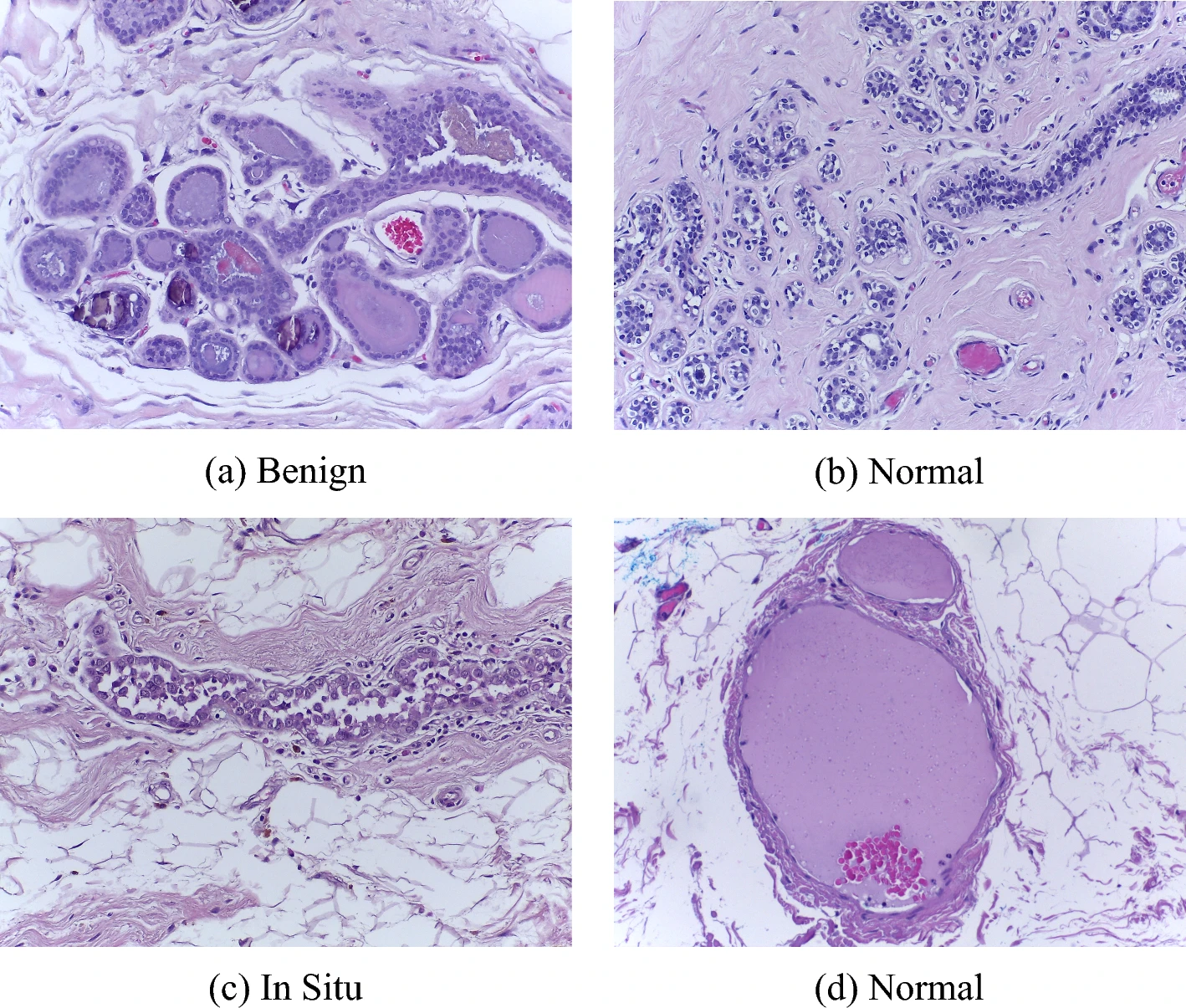
When a piece of suspicious breast tissue is removed, it goes to a lab, where it is cut into slices thin enough for light to pass through, mounted on a glass slide, and stained with dyes that make cell structures visible. A pathologist examines it under a microscope and decides whether the tissue is normal, whether the change is harmless, or whether it is cancer, and if so which kind. Many labs now scan those slides into digital images, which makes them readable by software.
That is the raw material here: not mammograms or MRI scans, but pictures of tissue at the cellular level, taken after a biopsy. This examination remains the decisive step in a breast cancer diagnosis. It is also slow, needs a trained specialist, and involves judgment calls. In one of the two image collections this study used, every image was labeled by two pathologists, and anything they disagreed on was thrown out — a practical admission that difficult slides can be read two ways.
Breast cancer is the most common cancer in women worldwide; the World Health Organization’s cancer agency estimated about 2.3 million new cases and about 666,000 deaths globally in 2022. Anything that helps a pathologist work faster, or miss less, has a large potential market.
The engineering problem is size
Plenty of AI systems already read tissue images well. The catch is that the strongest ones are big. They need powerful graphics processors, which means either costly hardware on site or sending patient images out to a remote data center.
So the team went the other way. Instead of one large model, they took two deliberately small ones — SqueezeNet and ShuffleNetV2, the second of which was built to run efficiently on phones and other low-power devices — and wired them together.
The wiring is the actual invention. Both networks look at the same image, and each produces its own set of observations. A component the authors call reciprocal cooperative gating sits between them and lets each network influence what the other pays attention to: signals the two networks merely duplicate are turned down, while signals that add something new are turned up. The merged result then goes to a final stage that produces the answer.
What the tests showed
The team used two public image collections that AI groups treat as reference sets for comparing systems.
The first, BreaKHis, was assembled at a single Brazilian laboratory from tissue samples of 82 patients. Here the model faced one question: benign or malignant? Working on images taken at one standard level of magnification (100×), it scored 99.72% on a test set of 352 images, which works out to a single wrong answer.
The second collection comes from a 2018 research competition in Portugal, and it is the one with the double-labeled images. It set a harder task: sort each image into four groups — normal tissue, a harmless change, cancer still confined to the spot where it began (in situ carcinoma), and cancer that has begun pushing into surrounding tissue (invasive carcinoma). Accuracy there was 97%, rising to 99% when the same collection was reduced to the simpler cancer-or-not question.
The researchers also ran statistical tests indicating that the gain over either network working alone was not a matter of chance, and published their code so others can repeat the experiments. They disclose that the work was supported by Russia’s Ministry of Economic Development. Dmitrii Kaplun of Innopolis University, one of the authors, has said in Russian-language comments that the two-network design suits regional hospitals and mobile devices.
What the number leaves out
The sharpest critic of these results is the paper itself.
AI systems are graded by showing them material they were not trained on. The Brazilian collection holds many images per patient, and the team divided images into training and testing groups without keeping any one patient entirely on one side. So the model may have been examined on tissue from patients it had already studied: a partly open-book exam. The authors state that this could make the result look better than it is, that splitting the data by patient would be the stricter and more realistic test, and that they intend to run it that way in future work. The researchers who built the collection a decade ago kept patients apart deliberately.
The test sets are also small. On the four-way task, 97% means 97 correct out of 100 images, so one extra mistake moves the headline figure by a full percentage point.
And these are reference collections, not clinics. The Brazilian images come from one laboratory; the Portuguese ones from patients in two regions of the country, all photographed through the same model of microscope and camera. In practice, slide preparation, staining chemistry and scanner models vary between labs, and models trained on one lab’s images often lose ground on another’s. At no point in this study did the model face images from a laboratory that contributed nothing to its training. There is also no comparison against how accurate pathologists themselves are, and no trial inside a working diagnostic service.
One gap matters especially for anyone weighing commercial prospects. In diagnostics, a missed cancer and a false alarm are not equivalent errors: one delays treatment, the other triggers unnecessary anxiety and procedures. The paper reports how often the model is right and how its mistakes split between the two kinds, but not how that balance shifts if the system is tuned to be more or less cautious. Hospital buyers and regulators look hard at exactly that trade-off.
The claim about small hospitals and mobile devices, similarly, follows from how the model is built rather than from a demonstration of it running on such equipment.
What comes next
The work still deserves attention. Two small models feeding off each other’s observations, instead of one expensive model, is a sensible response to a real constraint, and the researchers named the weakest part of their own testing rather than leaving it to be found.
But the distance between a benchmark score and a working product is long and well mapped. The steps here: retest with patients kept strictly apart, then with images from labs that contributed nothing to the training, then alongside pathologists in a working diagnostic service, with regulatory clearance after that. Each stage tends to trim the headline number.
For now, the accurate description is a promising laboratory result with a measurement flaw its own authors have flagged. That is a normal place for research to sit. It is also some way from a diagnosis anyone should lean on.